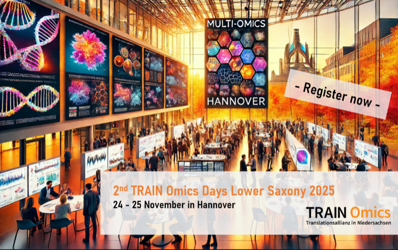
MoReHealth Niedersachsen is an innovative research project bringing the latest multi-omics research—meaning the simultaneous analysis and interpretation of different biological layers such as DNA, proteins, and metabolic products—into broadly applicable, personalized healthcare. The project is conducted by leading experts from top scientific institutions in Lower Saxony: Hannover Medical School (MHH), University Medical Center Göttingen (UMG), the Helmholtz Centre for Infection Research in Hannover, and the Technical University of Braunschweig. Specifically, the participating researchers investigate why older people are particularly susceptible to infectious diseases and how this knowledge can be used to better predict, prevent, and treat such illnesses. The aim is to leverage modern technologies to open up new opportunities in personalized prevention, diagnostics, and therapy.
MoReHealth receives four years of funding totaling 3 million Euros from the Lower Saxony Ministry of Science and Culture and the Volkswagen Foundation. Led by Prof. Thomas Illig (Hannover Unified Biobank, MHH), the project launches on 1 September 2025. The subproject at UMG is carried out at the Institute of Human Genetics and led by Prof. Bernd Wollnik and Dr. Julia Schmidt.
Case Study: Increased Risk of Infection in Older Age
As people age, their immune defenses decline, making older adults more vulnerable to infectious diseases such as shingles (Herpes zoster). While the illness can be mild, it often leads to serious complications involving the nervous system in older adults, sometimes resulting in chronic pain or neurological damage. Improving patient care requires a better understanding of the genetic and biological reasons behind these varied courses of disease.
The researchers link, analyze, and interpret data generated through genomics, transcriptomics, and metabolomics—including samples and data collected as part of the RESIST Excellence Cluster at Hannover Medical School. Thanks to technological advances, omics data can now be generated relatively quickly and inexpensively. However, the main challenge lies in analyzing and interpreting these massive datasets. MoReHealth develops standardized procedures for collecting, storing, and interpreting multi-omics data. This includes creating a secure, modular data platform and developing AI-driven tools to help decipher and better understand complex biological interactions.
Genomics at the Institute of Human Genetics, UMG
Genomic data are at the heart of the MoReHealth project. The team led by Prof. Bernd Wollnik, Director of the Institute of Human Genetics at the University Medical Center Göttingen (UMG), uses long-read genome sequencing to decode the complete DNA of 100 individuals from the RESIST cohort. This advanced technology can read very long stretches of DNA—spanning multiple genes and even entire chromosomes—allowing researchers to detect structural changes and rare genetic variants that traditional methods would miss. The goal is to identify genetic risk factors that increase susceptibility to viral infections or affect individual immune responses. Such findings are crucial for more accurate diagnostic procedures and truly personalized therapies.
Another key part of the project focuses on analyzing variants in mitochondrial DNA (mtDNA) using specialized sequencing techniques. Mitochondria, the “power plants” of the cell, have their own small genome. As we age, changes accumulate in mtDNA—a process increasingly linked to weakened immunity and chronic inflammation. The team of Prof. Wollnik and Dr. Schmidt investigates mtDNA mutation patterns that might predict poor outcomes in viral infections or lead to chronic complications. Their aim is to understand how these mutations impact cellular energy production and the immune system. In collaboration with experts at TU Braunschweig, they are also using state-of-the-art metabolomics technologies to explore the metabolic disturbances caused by mtDNA mutations.
A Strategic Investment in Precision Medicine
The significance of MoReHealth extends beyond this specific case study: The project pioneers a model for harnessing large-scale molecular data in health research—including the development of a scalable, GDPR-compliant multi-omics data platform that meets international quality standards and can be integrated seamlessly across different research institutions. By leveraging AI-driven analytical tools, MoReHealth aims to make multi-omics data actionable for clinical decision-making.
For Prof. Wollnik, MoReHealth is far more than a research project: “MoReHealth is a strategic investment in the future of personalized healthcare in Lower Saxony. With this pilot use case—age-related vulnerability to severe viral infections—we’re building a robust, validated, and transformative framework. It enables the use of multi-omics data in medicine to predict, prevent, and treat diseases based on each individual’s unique molecular profile. At the same time, MoReHealth sets new standards for sustainable, collaborative research infrastructure in Lower Saxony.”
Contact: Prof. Dr. med. Bernd Wollnik, Institute of Human Genetics, bernd.wollnik@med.uni-goettingen.de


